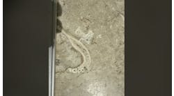
A 77-year-old female presents for a new-patient exam. Clinical assessment reveals two irregularly shaped radiopaque masses on the right posterior angle of the mandible.

Editor's note: This article first appeared in DE's Breakthrough Clinical with Stacey Simmons, DDS. Find out more about the clinical specialties newsletter created just for dentists, and subscribe here.
Presentation and chief complaint
A 77-year-old female presents to the office for a new-patient exam. Her chief complaint was that she was due for a cleaning and check-up.
Health history
Health history included cholesterol and blood thinner medications, type 2 diabetes, history of chronic obstructive pulmonary disease (COPD), high blood pressure, and sensitivity to penicillin.
Clinical exam
Assessment of the panoramic radiograph revealed two corrugated, irregularly shaped radiopaque masses on the right posterior angle of the mandible, each measuring approximately one inch in length. The area was not tender to palpation, and the patient had no knowledge that the lesions were there.
What are your differentials and subsequent recommended treatment modalities? Send your answers to [email protected]. Next month, we will present the final diagnosis and recommended treatment for this case.
CALL FOR PATHOLOGY CASES:
Do you have an interesting oral pathology case you would like to share with Breakthrough’s readers? If so, submit a clinical radiograph or high-resolution photograph, a patient history, diagnosis, and treatment rendered to [email protected].
Editor's note: This article first appeared in DE's Breakthrough Clinical with Stacey Simmons, DDS. Find out more about the clinical specialties newsletter created just for dentists, and subscribe here.