Clinicians of any specialty may identify oral pathology during clinical exams, so keeping your detection skills sharp is essential. One way to improve your clinical knowledge is to learn from cases your colleagues have seen. Use these clinical presentations and diagnoses from DentistryIQ readers to help determine treatment in your future cases. This can be an excellent reference for your practice.
Patient: 13-year-old female
- Large, raised lesion (1.5 cm) on the right dorsal and lateral aspects of the tongue, extending to the underside
- Lesion normal in color and slightly firmer than the surrounding area
- Not tender to palpation and no inflammation
- Tenderness upon mastication due to size of lesion
Patient: 67-year-old male
- Chief complaint: “scab on my lower lip for about eight weeks”
- Patient says lesion bleeds occasionally when eating, but reports little pain in the area
- Round lip lesion (1.6 cm in diameter) with an encrusted center that is firm and asymptomatic to palpation
- No cervical lymphadenopathy detected
A lesion suspicious for lip cancer
Patient: 62-year-old male
- Chief complaint: severe burning feeling on tongue and insides of cheeks, accompanied by “white patches all over”
- Patient has recent dry mouth, type 2 diabetes, high blood pressure
- White leukoplakic patches on right and left lateral borders of the tongue and on the buccal mucosa throughout
- A swipe with gauze removes the lesions
Severe burning and white patches
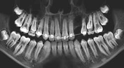
Patients: 11-year-old male and 14-year-old male
- Atypical eruption patterns, primarily for the second molars in the 14-year-old
- Large radiopacities around the second molars on the right side in both patients
- No pain upon palpation
Molars with irregular eruption patterns
Read more pathology cases …
Readers: Have you recently encountered a pathology case in your practice that you’d like to share? Email your details to Bethany Montoya, BAS, RDH, the editorial director of Clinical Insights. We look forward to featuring you!
Editor’s note: This article first appeared in Clinical Insights newsletter, a publication of the Endeavor Business Media Dental Group. Read more articles and subscribe.
About the Author
Vicki Cheeseman
Associate Editor
Vicki Cheeseman is an associate editor in Endeavor Business Media’s Dental Group. She edits for Dental Economics, RDH, DentistryIQ, and Perio-Implant Advisory. She has a BS in mathematics and a minor in computer science. Early on she traded numbers for words and has been happy ever since. Vicki began her career with Dental Economics in 1987 and has been fascinated with how much media production has changed through the years, yet editorial integrity remains the goal. In her spare time, you’ll find her curled up with a book—editor by day, reader always.


