I almost missed this lesion on the radiograph
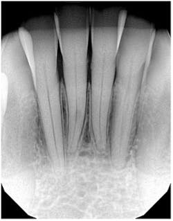
The distinct radiolucency on no. 25, just below the lower apical half of the tooth, is external resorption. The key diagnostic feature that differentiates it from internal resorption is the fact that the canal is defined. The CBCT confirms this, of course.
The patient presented asymptomatic (as these lesions usually are), and after discussion, we moved forward with a referral for assessment of extent and considerations for anticipated replacement.
External and internal resorption are some of the vaguest, yet greatest, topics of interest we have in dentistry. Their genesis, how they “behave,” and treatment modalities are indeed a conundrum. A refresher is always warranted. Take a look at these two articles (links below) that I’ve written on this topic. They’re worth a read or at least a quick scan.
- Endodontics reference guide: Distinguishing differences between internal and external resorption
- Internal resorption: A brief review and case report
Editor’s note: This article first appeared in Through the Loupes newsletter, a publication of the Endeavor Business Media Dental Group. Read more articles and subscribe to Through the Loupes.
About the Author

Stacey L. Gividen, DDS
Stacey L. Gividen, DDS, a graduate of Marquette University School of Dentistry, is in private practice in Montana. She is a guest lecturer at the University of Montana in the Anatomy and Physiology Department. Dr. Gividen has contributed to DentistryIQ, Perio-Implant Advisory, and Dental Economics. You may contact her at [email protected].