Cone Beam Fo

News you can use: Safe use of cone-beam CT; a topical gingival patch containing natural extracts

Aug. 17, 2012
Maria Perno Goldie, RDH, MS, reports on the recommendations for the safe use of cone-beam CT in a dental practice released by the American Dental Association Council on Scientific Affairs, and discusses the conclusions from a study involving a topical gingival patch containing natural extracts used to treat gingival inflammation.
Safe use of cone-beam CT
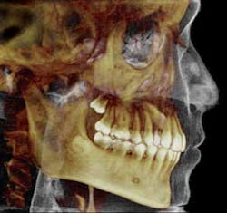
The American Dental Association (ADA) Council on Scientific Affairs (CSA) released recommendations for the safe use of cone-beam CT in a dental practice.(1,2,3) The CSA promotes safe use of conebeam computed tomography (CBCT) and appropriate professional justification of CBCT imaging procedures.
CBCT imaging provides three-dimensional volumetric data construction of dental and associated maxillofacial structures with isotropic resolution and high dimensional accuracy. The CSA reviewed the current research literature to develop collaborative guidance regarding the use of CBCT in dentistry with input from a broad group of stakeholder organizations. The Council’s principles for CBCT safety may be applied to any number of potential dental CBCT imaging applications. These devices were introduced to the market in the U.S. in 2001. This may have implications for many dental practices. As with other radiographic modalities, CBCT imaging should be used only after a review of the patient’s health, risk assessment, imaging history, and the completion of a thorough clinical examination. Dental practitioners should prescribe CBCT imaging only when they expect that the diagnostic yield will benefit patient care, enhance patient safety, or improve clinical outcomes significantly. In accordance with the National Council on Radiation Protection & Measurements’ (NCRP’s) Report No. 145 and standard selection criteria for dental radiographs, clinicians should perform radiographic imaging, including CBCT, only after professional justification that the potential clinical benefits will outweigh the risks associated with exposure to ionizing radiation.(4,5) Several organizations have released position statements and professional guidelines for cone-beam CT use, including the American Academy of Oral and Maxillofacial Radiology (AAOMR), the International Congress of Oral Implantologists, and the American Association of Endodontists.(6,7) For the ADA advisory statement, the council reviewed the current science, guidance, and other resources from professional organization, and also sought input from a number of stakeholder organizations, including the AAOMR, the American Academy of Oral and Maxillofacial Pathology, the American Academy of Pediatric Dentistry, the American Academy of Periodontology, the National Council on Radiation Protection and Measurements, and the U.S. Food and Drug Administration.References 1. Journal of the American Dental Association, August 2012, Vol. 143:8, pp. 899-902. 2. Updated information and services, including high-resolution figures, can be found in the online version of this article at: http://jada.ada.org/content/143/8/899. 3. This article cites 8 articles, 2 of which can be accessed free:http://jada.ada.org/content/143/8/899/#BIBL. 4. National Council on Radiation Protection & Measurements. Radiation Protection in Dentistry (Report No. 145). Bethesda, Md.: NRCP Publications; 2003. 5. American Dental Association; U.S. Department of Health and Human Services. The Selection of Patients for Dental Radiographic Examinations. Chicago: American Dental Association; 2004. www.ada.org. 6. Joint Position Statement of the American Association of Endodontists and the American Academy of Oral and Maxillofacial Radiology. USE OF CONE-BEAM COMPUTED TOMOGRAPHY IN ENDODONTICS. http://www.aaomr.org/resource/resmgr/Docs/AAOMR-AAE_postition_paper_CB.pdf. 7. Benavides E, Rios HF, Ganz SD, An C-H, Resnik R, Reardon GT, Feldman SJ, Mah JK, Hatcher D, Kim M-J, Sohn D-S, Palti A, Perel ML, Judy KWM, Misch CE, and Wang H-L. Use of Cone Beam Computed Tomography in Implant Dentistry: The International Congress of Oral Implantologists Consensus Report. Implant Dentistry, Vol 21:2, p 78-86.A topical gingival patch containing natural extracts Gingival inflammation often continues despite the use of conventional therapies. A topical gingival patch containing natural extracts has been shown in a randomized clinical trial to reduce gingival inflammation when administered as an independent therapy.
There was a case series of patients with moderate-to-severe chronic periodontitis in whom the patch was used together with scaling and root planing (SRP).(1)

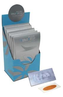
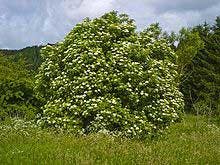
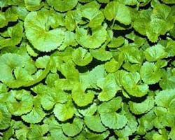
The researchers used a commercially available topical patch (PerioPatch) that contains extracts of three herbs: Centella asiatica; Echinacea purpurea; and Sambucus nigrad, that have all been shown to be both safe and effective in reducing gingival inflammation.(2,3,4,5)

These herbs, in combination with other formulatory agents in the patch, absorb inflammatory fluids and surface exudates from inflamed tissue as well. The patch is a registered medical device and was developed to help reduce localized irritation and inflamed gingiva.(6)

A small study following a series of 20 healthy patients, who presented with moderate-to-severe chronic periodontitis (probing depths, 5 to 8 mm), were treated with the patch immediately after treatment with SRP. All patients had bilateral gingival inflammation with a gingival index (GI) ≥ 2 and bleeding on probing (BOP) on ≥ 1site per tooth on two adjacent teeth.

After SRP, the patch was applied first by the study periodontist (AS), with two subsequent applications over the next 24 hours by the patient, and reevaluated two (2) to four (4) weeks later. On a separate visit, the contra lateral side was treated with SRP alone and was also reassessed after two (2) to four (4) weeks. Both the patch and non-patch sides showed improvement after SRP. When comparing the two sides, the patch-treated area showed a significantly greater reduction of inflammation and bleeding, as measured by GI (P <0.001) and BOP (P <0.002).

The conclusions of the study was, in this case series, that the patch was shown to provide a beneficial effect by helping to further reduce inflammation when used in conjunction with scaling and root planing. Additional research of the patch combined with SRP treatment is warranted within the framework of large randomized and controlled trials.

Visit the PerioPatch website to see a video of how the product works.(7)

References
1. Saffer A and Samuels N. A Novel Adjuvant Treatment to Scaling and Root Planing With a Topical. Gingival Patch: A Case Series. Clin Adv Periodontics 2012;2:123-127.
2. Sastravaha G, Yotnuengnit P, Booncong P, Sangtherapitikul P. Adjunctive periodontal treatment with Centella asiatica and Punica granatum extracts. A preliminary study. J Int Acad Periodontol 2003;5:106-115.
3. Sastravaha G, Gassmann G, Sangtherapitikul P, Grimm WD. Adjunctive periodontal treatment with Centella asiatica and Punica granatum extracts in supportive periodontal therapy. J Int Acad Periodontol 2005;7:70-79.
4 . Gasiorowska I, Patalas B, Wiktorowska B. Echinacin externa use in the treatment of periodontopathies (in Polish). Czas Stomatol 1981;34: 677-679.
5. Harokopakis E, Albzreh MH, Haase EM, Scannapieco FA, Hajishengallis G. Inhibition of proinflammatory activities of major periodontal pathogens by aqueous extracts from elder flower (Sambucus nigra). J Periodontol 2006;77:271-279.
6. PerioPatch, Izun Pharmaceuticals, New York, NY. http://www.perizoneonline.com/tag/periopatch.
7. http://www.perizoneonline.com/periopatch-how-it-works.

Note: PerioPatch is a product of Izun Pharmaceuticals, New York, New York. The study drug was provided free of charge by Izun Pharma, Jerusalem, Israel, which funded the statistical analysis of the data. Dr. Saffer is Senior Vice President for research at Izun Pharma, Jerusalem, Israel.

Maria Perno Goldie, RDH, MS

To read previous RDH eVillage FOCUS articles by Maria Perno Goldie, go to articles.