To see is to diagnose
By Howard Rothschild, DDS, FACD
What you cannot see, you can neither diagnose nor treat. In many practices today, what the patient cannot see or feel, you cannot treat. DIFOTI allows the dentist not only to make an early diagnosis of many dental problems, but also have the ability to visually demonstrate his or her concern to the patient. You can actually see a change take place in the patient's expression after they have seen the DIFOTI image and realize that he or she owns the problem. The patient is particularly pleased that a DIFOTI examination is radiation-free.
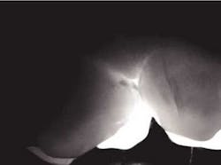
Digital Imaging Fiber-Optic Trans-Illumination (DIFOTI) is a diagnostic system for early detection of tooth decay and other changes in coronal tooth anatomy, including breakdown of restorative materials. It is a simple, painless, non-radiation, non-invasive procedure that can be repeatedly used with no risk to the patient. It creates real-time images on a computer monitor and stores them in a patient database on a PC. A small beam of light is passed through the contact area of adjacent teeth. If a carious lesion is present, the alteration of the enamel deflects the light beam producing a shadow.
null
Early inter-proximal decay #12 (DIFOTI and X-ray)
null
Peers, et al in 1993 found that the sensitivity of fiber-optic transillumination in the discovery of dental caries exceeded that of radiographs. Early phases of tooth decay are currently difficult to detect. While radiographs can disclose established cavities, particularly those that occur between the teeth, they are not effective in detecting early decay. Vaarkamp and Veerdonschot demonstrated that early diagnosis of approximal carious lesions was feasible when light was propagated through the carious tissue.
Schneiderman, et. al. (Caries Research 1997) demonstrated that DIFOTI is two times more sensitive than bitewing radiographs for detection of interproximal tooth decay (X-ray 31 percent; DIFOTI 69 percent); four times more sensitive for occlusal decay (X-ray 20 percent; DIFOTI 80 percent); and 10 times more sensitive for smooth surface decay (X-ray 4 percent; DIFOTI 41 percent).
Accurate diagnosis of occlusal caries is problematic at best. Prior to fluoride, the explorer could be retained in an occlusal fissure by factors other than decay. Brown in The Journal of Dental Education (1993) had concerns relative to the reliability and validity of making a diagnosis of occlusal decay based on the retention of an explorer in an occlusal fissure.
By the time there is radiographic evidence of decay, much of the occlusal enamel is undermined. Occlusal decay in patients who have used fluoride in the early years of life has a different anatomical model. The fissures are not open, thus do not give easy access to the dental explorer. The carious lesion starts at the base of the fissure and spreads within the tooth. DIFOTI is particularly efficient in diagnosing this type of decay.
null
Many may say the above DIFOTI image (Fig. 2) only demonstrates stained fissures. The explorer would not enter the fissures at all. The DIFOTI diagnosis was confirmed with a Diagnodent reading in excess of 70. The tooth was restored in a minimally invasive fashion.
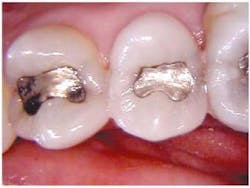
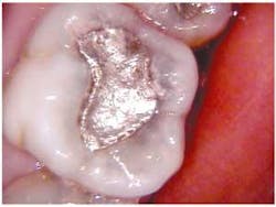
Recurrent caries is difficult to diagnose around an existing amalgam using radiographs. An occlusal DIFOTI image (Fig. 3) will demonstrate recurrent caries around an existing dental restoration.
Early anterior inter-proximal caries is easily diagnosed with DIFOTI (Fig. 3, below) even though the X-rays are essentially negative.
null
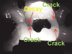
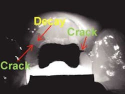
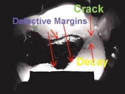
Interproximal vertical fractures emanating from existing amalgam restorations are easily seen and demonstrated to the patient. This case not only demonstrates deep vertical cracks but also decay.
In Fig. 4 (below) teeth numbers 3, 4, and 5 are shown. It would be difficult to demonstrate to the patient the problems that are present.
null
null
null
null
null
In the three DIFOTI images which begin the next column (Fig. 5), vertical fracture lines and decay shadow can easily be seen by the doctor and patient. The final image (Fig. 6) is of the teeth restored with CEREC porcelain inlays.
When a patient has recurrent pain in an endodontically treated tooth a fracture may be the cause. DIFOTI can easily demonstrate internal vertical fractures.
null
DIFOTI can be used as an adjunctive instrument in your routine examination. Photographic documentation from DIFOTI images is very useful when submitting insurance claims. When used routinely, the dentist will become more and more dependent on DIFOTI. For the dental economists reading this article, ROI is between two to three months.
Dr. Howard Rothschild has a practice focusing on cosmetic, restorative, and implant dentistry in Baltimore, Md. Dr. Rothschild can be reached at (410) 602-8100.

