Lava Case Study
Introduction:
Despite all the advancements in porcelain bonding technology, the ideal aesthetic bridge material has remained elusive. The first characteristic of this material would be translucency, which would allow light to pass through the material, thus creating the illusion of real enamel. Translucency also is important for lighting up the gingiva. Crowns and bridges with metal cores do not meet this criterion because they prevent light from reaching the roots. This, in turn, prevents the fiber-optic back-lighting effect seen with natural teeth. Instead, the metal core restorations cast a shadow on the gingiva, causing what should normally look like pink, healthy tissue to appear gray and lifeless or sometimes inflamed. This phenomenon also occurs to a lesser extent in some all-ceramic restorations, which are fabricated on very opaque core materials. The ideal restorative substructure would have the optical qualities of dentin, thus allowing uninterrupted transfer of light throughout the tooth.
Secondly, the ideal bridge framework would be very strong so that the bridge connectors could be made as small as the contacts between real teeth simulating deep labial embrasures. This would allow the teeth natural-looking separation. Up until now ceramic frameworks have required a connector size greater than 9 mm2, which typically creates an unnaturally long contact with a shallow embrasure when placed between average-sized anterior teeth. Also, the ideal framework would have an excellent fit and be simple to develop. It would be compatible with porcelain, so that the bond strength between the porcelain and the framework would be strong enough to withstand heavy biting forces.
Another concern with metal is the depth of reduction necessary to develop color. Even in the hands of the most skilled technicians, a reduction of less than 1.4 mm allows the opaque layer of metal underneath to show through the porcelain. This amount of reduction is safe on a molar or husky cuspid, but is immoderately aggressive for a lateral incisor or thinner teeth and could compromise their long-term prognosis. This means that where the reduction for aesthetics is the most critical, it also is the most potentially destructive to the dentition.
To remedy this complication, the suggestion has been made by some laboratory technicians and dental practitioners to cut back the metal framework inside the porcelain so that light can pass through on the facial surface of the bridge abutment. However, this leaves the clinician with the dilemma of cementing the bridge with conventional cement, which provides a weak juncture between the porcelain and the tooth, allowing the potential for fracture. Conversely, if the bridge is cemented with bonding resin, the porcelain will have a strong bond, but one that may be stronger than the metal itself. If any micromovement such as flecture occurs in the tooth, the bonded porcelain below the cutback on the metal framework would likely fracture at the metal margin.
The introduction of zirconium oxide as a restorative material satisfies some of our aesthetic and functional needs. The higher flexural strength of this material allows smaller, more aesthetic bridge connectors to be used. Light passes through the zirconia core material giving some illumination of gingival tissues. The amount of light transmission varies greatly from manufacturer to manufacturer, as does the thickness of the core material. Aesthetic results improve as the core materials become thinner, but strength as well as aesthetic requirements must be considered.
3M™ ESPE™'s Lava™ All-Ceramic System core material comes close to the optical qualities of dentin. Because the marginal areas involve the enamel layer, for optimal results the marginal areas should be thinned significantly to replicate the light transmission of enamel (.2 to .3 mm); or the core material can be cut back from the margin and a traditional porcelain butt joint can be employed.
Experimentation with other types of metal hybrid bridges in which the metal is surrounded with porcelain has demonstrated some success. Variations of the technique which totally encapsulate the metal within a pressed ceramic matrix have been successful to date but lack long-term clinical testing. However, in the following case study, the potential simplicity and aesthetics of layering the porcelain over a strong, dentin-colored framework outweigh the benefits of these metal hybrid bridges.
CASE STUDY:
Patient history —
A healthy, 38-year-old female expressed a desire to replace her missing teeth, especially the upper-left bicuspid because the gap was visible when she smiled. The patient worked with the public and wanted to be able to smile with confidence. Tooth No. 13 had been removed seven years ago. The patient had been managing the space with a temporary, removable partial denture, which had recently fractured. Tired of wearing the appliance, the patient sought a new replacement option. The patient did not want any metal in her mouth for both aesthetic and perceived health concerns.
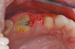
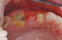
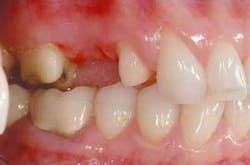
The patient's teeth were generally healthy. Excellent oral hygiene was evident, and there was virtually no active decay, other than an open margin on the five surface amalgam restoration on Tooth No. 14. Although the patient had no signs of any inflammation or periodontal disease, there were localized moderate recessions of 1.5 mm on Tooth No. 12 and No. 14. The patient admitted to having a clenching habit, but presented without any TMJ symptoms. Further, the patient's occlusion was generally stable except for drifting molars — Tooth No. 31 and No. 32, on the right side. By replacing Tooth No. 13, the patient would have a stable occlusion from No. 5 to No. 15 on the maxillary arch, and No. 18 to No. 28 on the mandibular arch (Tooth No. 29 and No. 30 were missing). For financial reasons, the patient wanted to wait to treat the lower right quadrant, which had been treatment-planned for molar uprighting and implants.
Treatment plan —
The patient was presented with an option for an implant and ridge augmentation for Tooth No. 13 and a bonded porcelain restoration for No. 14 to replace the failing amalgam. She declined. The patient expressed a desire for a bridge that looked like her natural teeth and contained no metal. Chief objections to the implant were the cost and time involved with the two proposed surgeries.
Tooth No. 14 had been previously restored with a five-surface amalgam. The buccal cusps were undermined with supragingival cracks. Further, there was recurrent caries, starting at the gingival margin on the distal. Tooth No. 12 was a virgin premolar, and it was decided to sacrifice the integrity of this tooth for what appeared to be a predictable abutment for a three-unit bridge.
Technique —
After a thorough examination, diagnostic models and bite registration were taken for a diagnostic wax-up of the bridge from Tooth No. 12 to No. 14. The crowns were waxed to the cementoenamel juncture (CEJ) on the facial surface and 1 mm coronal to the gingival margin on the lingual surface. The laboratory was instructed to keep the wax away from the gingival 1 mm as a time-saver. When the matrix is filled with the temporary composite resin, the tight-fitting gingival margin makes a clean cut for easy trimming. The provisional (prototype) bridge matrix was formed by molding Sil-tech® putty (Ivoclar Vivadent) over the diagnostic wax-ups. A vacuum-formed matrix for measuring the depth of reduction was formed on a duplicate model of the wax-ups. This matrix also can serve as a backup if something should happen to the wax-ups or the Sil-tech putty matrix.
One of the main reasons for this extra step with the reduction guide is that restored teeth may require some change from their existing to their desired final contours. In this case, a fuller buccal corridor was desired and a small amount of extra wax was placed on the facial surface of Tooth No. 12 and No. 13 to fill out the smile. This, in turn, decreased the overall amount of tooth reduction necessary on the buccal surfaces of the teeth to achieve adequate space for the framework and the porcelain.
null
null
null
null
null
null
It was decided to treat the bridge without any tissue grafting because the bridge was located in an area of less aesthetic concern than the anterior teeth. The loss of volume in the ridge was minimal. With the wax-up, it was determined that an aesthetic result could be achieved without a graft if a ridge-lap pontic was used. The patient does want to pursue connective tissue grafting over the recessions on Tooth No. 12 and No. 14 at some future date. For this reason, it was decided to end the margins of the bridge in the same place where the CEJ would ideally be seen. This can serve as a surgical guide for the positioning of the graft when it is completed.
On the preparation day, the patient was anesthetized with one carpule of 2 percent Septocaine. Both bridge abutments were prepared the same way with 1.5 mm occlusal reduction prepared with a KS 2 coarse 1.4 mm round-tipped, straight diamond bur (Brasseler) and a coarse diamond football (Brasseler). The facial and lingual surfaces were prepared with a KS 2 coarse, followed by a KS 1 fine for smoothing and final contouring. The inter-proximal contacts were prepared with a KS 0 coarse 1.0 mm round-tipped, straight diamond bur (Brasseler). Although crack propagation may not be a concern with the Lava All-Ceramic System, which was utilized to develop the bridge, still prepare the teeth to be very smooth with no sharp angles to potentially propagate any cracks. The depth of reduction in the photos may seem minimal based on the shape of the original teeth. However, because the plan in the wax-up was to fill out the buccal corridor, the depth cuts were based on a thickness determined by the final wax contour, not the original tooth contour.
In this case, no retraction cord was necessary because of the supragingival margins. An impression was taken with 3M ESPE's Impregum™ and Permadyne™ impression materials.
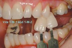
The impression, opposing models, two matching bite registrations, and shade tab photos were sent to Matt Roberts at CMR Dental Lab in Idaho Falls, Idaho, with instructions to make a Lava All-Ceramic System bridge with a ridge-lap pontic.
Stone casts were made from the impressions and mounted on an Artex (Jensen Industries) articulator with the face-bow and bite registrations provided. Margin trimming of dies is conducted in a way that leaves very distinct margins that are easily readable for the laser scanner employed by the Lava All-Ceramic System. Articulated models with the trimmed dies were sent to 3M ESPE for the fabrication of the Lava All-Ceramic System substructure.
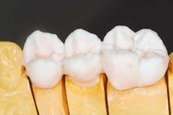
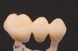
When the framework was returned to the laboratory, it was examined for fit on a master model. Initial fit was good. Small adjustments in angulations of the emergence profile were made and the facial margins were cut back for porcelain margins to increase gingival illumination. Although a ridge-lap pontic was to be utilized, the stone was relieved enough to create pontic pressure against the tissue and ensure the development of a small gingival cuff, creating the illusion of natural emergence.
The ceramic build-up was started with the application of the porcelain margin material and some highly chromatic modifiers to camouflage the substructure. These layers were fired under vacuum in a porcelain furnace to full maturity. Subsequent layers of dentin and enamel powders were built and fired to replicate the optical qualities of the patient's natural dentition. Anatomical features were built in with a porcelain brush, rather than ground in with a high-speed handpiece, to create more natural contours. Final surface texture was achieved by grinding with a 850-016 diamond bur (Brasseler), then the bridge was fired to a low glaze. A diamond-impregnated rubber polishing wheel (Brasseler) was used to smooth areas to be highlighted, and diamond polish (Brasseler) accomplished the final surface luster. The bridge was fit to a fresh solid model to verify the interproximal contacts, tissue adaptation, and occlusion, then returned to Dr. Jones for delivery.
At the framework seating appointment, the patient had only minimum sensitivity and chose not to be anesthetized. The temporary bridge was removed and the final Lava All-Ceramic System bridge was tried-in with warm water. The interproximal contacts were checked for tight or open contacts. The abutment preparation margins were checked with a 10X power surgical microscope and an explorer for any gaps or overhangs. The pontic was checked for tight tissue adaptation and the occlusion checked for even centric stops in the fossa and on marginal ridges. It was determined that there were no working or balancing interferences and no teeth out of occlusion on the bridge.
The bridge was rinsed and cleansed with Ultra-Etch® (Ultradent) phosphoric acid to remove proteins and contaminants, then rinsed with water and dried with air. The bridge abutments were cleansed with lab pumice and warm water on a prophy cup and a Benda®Brush Microfine (Centrix, Inc.) used for the margins, carefully avoiding the gingiva. The pumice residue was wiped away with PREP disinfectant (The Value Dental Company, Inc.). The assistant mixed 3M ESPE RelyX™ Unicem Self-Adhesive Universal Resin Cement, a self-etching resin cement, while the doctor dried the abutment teeth, carefully avoiding desiccation of the dentin. A thin film of cement was applied to the internal surface of the abutment crowns, covering all of the margins.
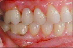
The bridge was seated and all of the excess cement was removed with the BendaBrush Microfine. (To ensure as easy a cleanup as possible with 3M ESPE RelyX Unicem Self-Adhesive Universal Resin Cement, be sure to remove all of the cement from the margins before it sets, because it bonds to any exposed enamel or dentin and can become difficult to remove. RelyX Unicem cement is a dual-curing cement that sets automatically in five minutes. It does not require any additional light-curing.) After cementation, the bridge and adjacent teeth were inspected for excess resin. The occlusion was confirmed before the patient was dismissed.
Conclusion
Utilizing 3M ESPE's Lava All-Ceramic System in combination with RelyX Unicem cement is an exceptional aesthetic technique for making three-unit bridges that are metal-free. Like any new technique, it will require evaluation over time in actual clinical practice to see if it lives up to its promise; however, today, Lava All-Ceramic System zirconia framework restorations offer a strong, aesthetic, all-ceramic restorative option for crowns and bridges. Further, because these restorations are cementable, as opposed to bondable, a wide variety of less technique-sensitive options, such as second molars and subgingival margins, exist. The next step will be an all-ceramic framework that can be used in a partial coverage abutment design. In the meantime, Lava All-Ceramic System bridges are an excellent choice for aesthetically sensitive and health-conscious patients who desire a metal-free, fixed restoration for missing teeth.
For further reference, consult 3M ESPE's Lava All-Ceramic System Technical Product Profile at www.3MESPE.com/labproducts.
Lynn Jones, DDS Dr. Jones is recognized for her excellent cosmetic dentistry and innovative management practices. She is an accreditation examiner for the AACD and lectures extensively on micro-dentistry. Director of "Aesthetics Continuums" for the University of Washington CDE, she maintains a private practice in Bellevue, Wash.
Matt Roberts Mr. Roberts, a highly recognized dental ceramist, is an accredited member of the AACD. He lectures nationally and internationally and has worked with leading clinicians around the world. He teaches advanced level laboratory and clinical programs for dentists and ceramists through Team Aesthetic Seminars.
