Using digital photography to put your dental treatment case acceptance rate on steroids!
A picture is worth a thousand words when it comes to case acceptance in dentistry. Dr. Gregory Wych details the series of photos he uses for case presentations and explains how his team incorporates digital photography into the practice schedule to help patients become engaged and excited about their dental treatment.
This article first appeared in the newsletter, DE's Breakthrough Clinical with Stacey Simmons, DDS. Subscribe here.
We all know the saying “A picture is worth a thousand words.” Nothing is truer than when it comes to digital photography and dental treatment case acceptance.
Intraoral photography was first developed in the 1980s. Primarily used in the dental hygiene operatories, showing pictures of teeth to patients was a rudimentary way to increase awareness and hopefully case acceptance. Certainly, for single-tooth dentistry and quick reminders of diagnosed, uncompleted dental care, intraoral photography can be useful. A hygienist or dental assistant capturing an image to show a patient a problem tooth is a quick and easy tool in the hygiene operatory.
For years, hygiene and dental consultants have taught the “tour of the mouth” at new patient and recare appointments. Clinicians take a number of single-tooth pictures as they go from Nos. 1 through 32 and beat the poor patient into submission with an overwhelming volume of photos that show the mouth in disrepair.
In my experience, however, intraoral photography is limiting. After patients have seen several pictures of teeth, they tend to zone out and become disengaged or even agitated by the onslaught of single-tooth pictures showing cracks and worn restorations. “Enough,” they say, "enough!"
That's ancient technology!
In the early 2000s, digital photography began to displace 35 mm slide photography. With slide photography, we often took many images with the hope that at least some would be presentable enough to show the patient at a later date. Obviously, digital photography allows us instant feedback, easy editing, and immediate presentation to the patient. We also are able to manipulate the images, dropping in smiles from a library to allow patients to see expected treatment results even before the case has started.
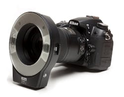
The type of camera that I use and recommend is an SLR-type camera, such as the one shown below (Nikon 7100, photomed.net.) This camera is lightweight, easy to use, and looks impressive to the patient.
Nikon 7100 camera
Several organizations, such as the American Academy of Cosmetic Dentistry (AACD) and continuing education institutes (such as Spear Education) teach advanced photographic techniques. The series of photos they advocate are excellent for documentation and treatment planning, but they tend to be overkill when used in treatment presentations to our patients.
The series of photos that I have had the most success with in case presentations is “The Magnificent Seven” (or eight or nine). The photos, in order of display, are:
- Full face
- Lip at rest
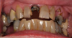
- Retracted full smile (with or without teeth separated)
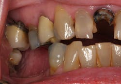
- Right lateral retracted
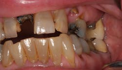
- Left lateral retracted
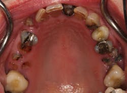
- Maxillary occlusal
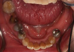
- Mandibular occlusal
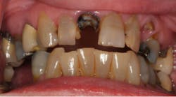
These are examples of the photographic series that we display to our patients:
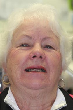
Full face
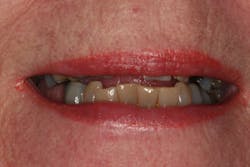
Lip at rest (If you are familiar with the concept of Facially Generated Treatment Planning, then you know the value of this particular photo and how to explain it to the patient.)
Retracted full smile
Right lateral view
Left lateral view
Maxillary occlusal
Mandibular occlusal
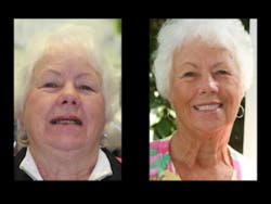
Finished result—before and after!
I have found that this series of photos will give patients all they need to see, own, and accept their dental care, yet not overwhelm them or be redundant with information.
In my office, we take photos at all new patient exams and for targeted hygiene recall appointments. During our morning huddle, when we are discussing the hygiene patients, we will plan to take photos of any patient who has diagnosed, uncompleted treatment and is due for hygiene recare radiographs (usually four bitewings and two anterior periapicals).
Choreography with the staff is the key to effective, efficient photography and presentation. None of the photography should be done by the dentist! Of course, having enough team members is mandatory. When a new patient is scheduled, the new patient coordinator first gives a tour of the office and then interviews the patient. When ready to begin gathering data on the patient, the coordinator will summon another assistant to help with the photos. While the new patient coordinator gathers the radiographic information (3-D scan, full-mouth series, etc.), the second assistant handles the photographic images. All of this is completed before the dentist enters the room for the new patient exam.
The photographic series is treated as just another part of “what we do around here.” The series takes about three to five minutes to complete. The second assistant then loads the images into our software, edits them, and arranges a standardized slide show for the dentist. We use an old image storage software called ImageFx, but there are many other image storage and editing software programs on the market, including PowerPoint, Keynote, Photodirector 8, and Paint Shop Pro 8.
Likewise, we target hygiene recare patients who have uncompleted treatment. At the beginning of the hygiene appointment, the hygienist summons an assistant to help take and load the digital photos, usually before the routine recare radiographs are taken. The process is simplified, because all of our team members wear walkie-talkies (Kisco Dental) for intraoffice communication. All data is gathered before the hygienist starts treatment and before the dentist does the hygiene recare exam.
Oversized monitors are mounted to the chairs in the treatment rooms. This gives patients an awesome view of their mouth. After the exam and reading the radiographs, I remove my mask, gloves, and loupes, sit next to the patient, and narrate exactly what I see as we review the images together. Patients become amazingly engaged with the photos and actually begin to diagnose themselves!
Many dentists think they are the only ones who can take the photos ... that they have to be perfect. However, team members can actually take better photos than the dentist, and with some simple editing software tools, the images turn out great. The photos just need to be good enough to show pathology.
The key is, patients don’t need to be lectured to; they need to get interested and engaged. When they become engaged in their photos, they own their disease. And when you show them photos of other patients who have accepted and completed care, they become excited and move forward with their own treatment.
We will often present the treatment plan at the initial new patient appointment. But when a case is large or if the patient has numerous options, we will invite the patient back for a treatment planning consultation. For these appointments, we print out the photos and give them to the patient (hopefully their significant other will be present also), along with a cosmetically enhanced image of their smile and the end result. We use a cosmetic imaging software called Envision A Smile, but there are others on the market (Snap, SmilePix, etc.). All of these tools help patients become engaged and excited about their treatment.
This is how you use digital photography to put your case acceptance rate on steroids!
This article first appeared in the newsletter, DE's Breakthrough Clinical with Stacey Simmons, DDS. Subscribe here.
