Endodontic case study: radix entomolaris
Clinician: Aneel Belani, DDS
Description of the tooth: Lower molar No. 19 of a 59-year-old female patient
Diagnosis: Irreversible pulpitis with normal apical tissue.
A 59-year-old female patient presented to our office for consultation on tooth No. 19 with the chief complaint: “I can’t drink anything cold or I have shooting pain in my tooth, and it’s giving me headaches.” Medical history was noncontributory. Patient had visited her dentist three weeks prior and had an amalgam restoration replaced with a bonded composite filling. She had returned to her dentist three days prior for an emergency visit and was referred to our office.
ALSO BY DR. ANEEL BELANI |Endodontic case study: Healing of a large lesion in a lower molar after proper dental instrumentation and irrigation
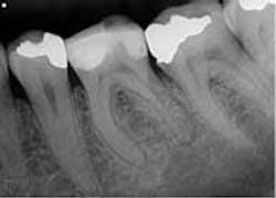
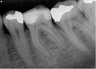
Clinical evaluation revealed no intraoral or extraoral swelling and a large composite restoration in tooth No. 19. Endodontic testing revealed severe pain to cold in tooth No. 19; all other tests were within normal limits (WNL). All other teeth in the quadrant tested WNL. The diagnosis was irreversible pulpitis with normal apical tissue. Radiographic exam revealed a large composite restoration and abnormal anatomy suggesting radix entomolaris. Radix entomolaris is a supernumerary root located distolingually in mandibular molars. The supernumerary root can also be found mesiobucally, and this is called radix paramolaris. The prevalence of a supernumerary root in mandibular molars has been found to be between 2.2% (Skidmore) and 21.5% (Yew and Chan). It is important to carefully study our preoperative and working radiographs in order to recognize the presence and location of abnormal anatomy.
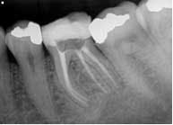
The patient was given treatment options and informed consent, and elected to begin treatment immediately. Profound anesthesia was achieved using two carpules of 2% lidocaine for IANB and one carpule of Septocaine for long buccal infiltration. The tooth was isolated with a rubber dam and accessed under Global operating microscope using round burs and an Endo-Z bur. The distal canal was located in its typical orientation, centered buccolingually between the mesial canals. However, the canal leading to the radix entomolaris was found far to the lingual and off-center. Working length was determined using Root ZX II electronic apex locator. ISO size 6, 8, and 10 files were used to create initial glide path. Instrumentation was completed using ProGlider files to establish glide path, and a combination between Protaper and Vortex Blue files with full-strength sodium hypochlorite irrigation. QMix was placed into the canals after irrigation and agitated with EndoActivator to remove the smear layer and for its antibacterial properties. The canals were filled using warm vertical condensation with System B and Calamus backfill. The patient was given a six-month follow-up appointment and ibuprofen 600 mg for postoperative discomfort.
Examples of supernumerary roots. Ribeiro and Consolaro (1997)
MORE ENDODONTIC CASE STUDIES …
Endodontic case study: referred pain leads to diagnostic uncertainty
Endodontic case study: an alternative method for the cleaning and shaping of a mid-mesial canal
Endodontic case study: Challenges of a bifurcated canal


