Editor's note: Originally published in December 2019. Updated February 14, 2022.
Case history
A 61-year-old male presented to my office for a sleep apnea consult. He doesn’t take any medications, has a history of five knee surgeries, two sinus surgeries, a ruptured kidney, five trigger finger surgeries, hernia surgery, carpal tunnel surgery, hand surgery for broken fingers, mumps, measles, and chicken pox.
Clinical assessment
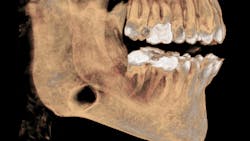
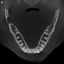
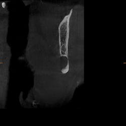
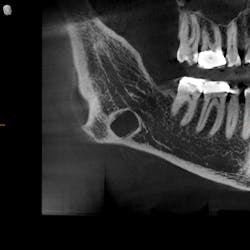
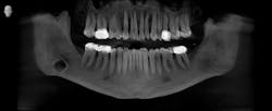
As part of our intake process to make a sleep mandibular advancement device, we take a cone beam image (figures 1–4). This way we can evaluate sinuses, septum, turbinates, teeth, and jaws.
Upon examination, it was noted that a dark, radiolucent lesion was present on the lower right jaw, anterior to the angle of the mandible. The patient didn’t have any pain, symptoms, or knowledge of the lesion. It was not palpable. He reported that he had his third molars removed in his twenties with no complications.
Differential diagnosis and discussion
Reflecting back on my amazing pathology education at Creighton University (thanks, Dr. Bob Achterberg!), I immediately tried to think of what my differentials could be. Full disclosure: I was worried that my patient’s jaw could break at any moment. I thought it was a residual cyst from third-molar removal.
The Stafne defect is code for a depression in the bone, a concavity, due to the submandibular gland. In addition, it can’t be qualified as a cyst because there is no epithelial lining or fluid content. It does usually occur more in men as well.1 There is no treatment necessary; it’s something we can just monitor.
Now, all I have to do is find some other defect so I can have something named after me!
Read another pathology case: An asymptomatic, fluctuant mass with a bluish hue
Reference
1. Hisatomi M, Munhoz L, Asaumi J, Arita ES. Stafne bone defects radiographic features in panoramic radiographs: assessment of 91 cases. Med Oral Patol Oral Cir Bucal. 2019;24(1):e12-e19. doi:10.4317/medoral.22592.
About the Author
Erin Elliott, DDS
Erin Elliott, DDS, a practicing dentist in Post Falls, Idaho, has successfully integrated dental sleep medicine into her practice. She lectures extensively and leads a hands-on workshop focusing on practical strategies for successful implementation of sleep medicine into the general practice. She is active in the American Academy of Sleep Medicine and American Academy of Dental Sleep Medicine, and she is a past president and diplomate of the American Sleep and Breathing Academy. Contact her at [email protected].
