Oral pathology case: Venous blue lake mole—what?
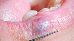
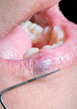
Taking care of the patient’s chief complaint was one thing, but what really caught my eye was an 8 mm x 8 mm purple/dark blue raised nodule on the right side of the vermilion border of her lower lip. There was no pain on palpation, and the nodule blanched somewhat when compressed (figure 2).
Since this was a new patient and because I’m one of those pathology hound dogs, I asked her about it. “Oh, that! It’s called a venous blue lake mole. I’ve had it looked at numerous times. My aunt has one on the same exact spot. I’ve been told that it’s harmless and that there’s nothing that can be done about it that won’t leave a large scar on my lip because of the size. I always try to cover it up with makeup and lipstick because I don’t like the look of it.”
Well, there ya go. Observation, diagnosis, and treatment all wrapped up in that short, sweet dialogue.
Now, I’ve seen pathology like this before and for the most part passed the nodules off as benign blood blisters or blue nevi lesions. And I suppose if I dug back far enough into the recesses of my brain, I could maybe remember the name venous blue lake mole from those pathology lectures in dental school. But let’s be honest, probably not. Nonetheless, this was a good lesson and refresher on pathology that we all likely see more often than not.
Just exactly what are venous blue lake moles? These lesions are not uncommon findings, especially in older generations who have spent some time in the sun. They range in size from 1 mm to 12 mm, and they slowly fill back up when compressed. Their etiology is from sun exposure that ultimately causes vascular damage.1 The ears, face, and lips are the most common locations for these lesions, and they can grow in size with trauma or continued insult.1,2 They are benign but can be confused with melanoma, so evaluation and follow-up are recommended.2 Typically, no treatment is needed unless it’s for cosmetic reasons. Surgical incision or laser therapy is effective for removal.
So, I’m sharing this because, come on, with a cool, legit name like venous blue lake mole, it’s definitely one to put in the memory bank to spew out over cocktails and impress your friends, right? Now you’re in the know. Good stuff.
If you don’t see any sort of lesion or pathology every day that warrants inquiry, referral, or biopsy, then you need to up your game. There is more pathology out there than we think. When you do find something, take a pic, share it with me, and I’ll pass it along. Refreshers on the common and not-so-common lesions are always useful.
References
- Venous lake. Wikipedia. Updated January 17, 2019. Accessed September 16, 2020. https://en.wikipedia.org/wiki/Venous_lake
- Venous lake. American Osteopathic College of Dermatology. Accessed September 16, 2020. https://www.aocd.org/page/VenousLake
Editor’s note: This article first appeared in Through the Loupes newsletter, a publication of the Endeavor Business Media Dental Group. Read more articles at this link and subscribe here.
More pathology cases:
About the Author

Stacey L. Gividen, DDS
Stacey L. Gividen, DDS, a graduate of Marquette University School of Dentistry, is in private practice in Montana. She is a guest lecturer at the University of Montana in the Anatomy and Physiology Department. Dr. Gividen has contributed to DentistryIQ, Perio-Implant Advisory, and Dental Economics. You may contact her at [email protected].