How Are Doctors Using CariVu? Find Out In This Post.
In my practice, I treat both fee-for-service patients as well as those who have dental insurance. However, even though caries detection is now covered under many insurance plans, I have not yet charged a fee for using this technology due to its clinical benefits.
I always strive to be a better diagnostician. With so many new dental products emerging each year, I'm always researching to find the best options to add to my diagnostic armamentarium. Recently, I implemented DEXIS
CariVu, a caries detection device that uses near-infrared transillumination technology.
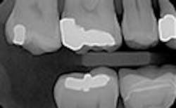
Patients often arrive at my office with poor-quality digital x-rays or printouts (figure 1) from their previous dentists and no caries detection images. I can't treat what I can't see, and that increases the potential for delayed diagnosis and treatment as well as missed income opportunities. In some cases, these poor-quality x-rays were taken so recently that it is not prudent to expose the patient to additional radiation. Having an alternative to radiological caries detection is a boon in these situations
For example, 18-year-old twins presented to my office recently. The referring practice was not able to supply clinically readable digital x-rays. To get the twins comfortable and start the diagnostic process, I used CariVu. I was able to determine that they did not have occlusal or interproximal caries, and consequently, they had no need for treatment at the time These young men remarked, "Dr. McKibben has all of the latest technology." They will probably even tell their friends and family. Although I am an older practitioner, I still want to maintain a reputation of being on the cutting edge of innovation. CariVu, along with my other technologies such as CAD/CAM, helps with that perception.
Here's another example of where a patient benefited from this technology's ability to accurately display interproximal caries.1 The patient needed a crown on tooth No. 3, and adjacent tooth No. 2 looked clean on a bitewing x-ray (figure 5).
Figure 5: Bitewing x-ray shows no indication of caries on tooth No. 2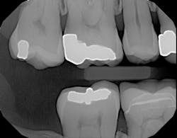
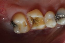
Before I began prepping for the crown, I used CariVu. On the images, I saw not only a crack on the tooth that was to be crowned, but also clear caries on the adjacent tooth (figure 6). During treatment, I snapped a picture of the preparation on tooth No. 2 with an SLR extraoral camera (figure 7). I took an additional photo mid-treatment to show how far the cavity had progressed into the dentin. The bitewing didn't show any of this decay. This caries detection technology gives me a great opportunity to tell patients that I found a cavity that we would not have known about until it caused trouble. There is no surprise later and no apologies for missing an early lesion.
Figure 6: CariVu image clearly shows mesial decay on tooth No. 2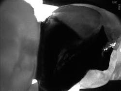
While some incipient lesions can be put on a "watch list," some should be treated early. Patients don't want to wait until they are in pain or having sensitivity, and that can be avoided by detecting and restoring caries early with smaller fillings that last longer and are less damaging to tooth structure. CariVu has given me another way to prove that I have confidence in my diagnoses. When I have shown x-rays to patients in the past, some have said, "I don't see it, but I trust you." I appreciate their trust, but what I really enjoy is being able to show proof of the caries, treat it, and then follow through.
Motivating patients by helping them understand the situation is an important part of treatment acceptance. The CariVu images appear like x-rays on my operatory monitors, look familiar to patients, and are very explanatory. The tooth appears light and the caries appears dark. My next investment for the practice is larger high-definition screens for my treatment rooms. The small screens are working fine, but with digital technology it is nice to be able to enlarge images as much as possible for diagnostics and patient education.
Whether you charge for caries detection, charge only for specific instances, or don't charge at all, this type of imaging method has many benefits. It increases patient education and case acceptance, and it gives me more information with which to recommend restorative treatment. Dr. Martin Garron, a mentor who encouraged me from childhood until his passing, once advised me about staying ahead of the competition by carefully adding effective technologies to my practice. He said, "Don't be the first, but don't be the last." Caries detection-especially the type that can show tooth structure that is undetectable on some x-rays-is a sensible diagnostic and financial addition to add to any practice as soon as possible.
Author's note: Dr. William McKibben has no financial interests in DEXIS, LLC. Indications for the use for CariVu can be found at dexis.com/ifu.
Reference
1. Kühnisch J. Benefits of the DIAGNOcam Procedure for the Detection and Diagnosis of Caries [study project]. Munich: Ludwig Maximilian University of Munich; 2013.
William J. McKibben, DDS, is a 1978 graduate of Georgetown University in Washington, DC. He is a member of numerous national dental organizations and is on the faculty of Esthetic Professionals of Tarzana, California. Dr. McKibben has completed residencies in advanced occlusion, advanced fixed prosthodontics, and comprehensive esthetics. He is currently in private practice in Long Beach, California.