Bone health, systemic health, and dentistry: Part 1
Key Highlights
- Osteoporosis risk begins early in life, making adolescent bone development and peak bone mass critical to preventing fractures later.
- Because osteoporosis often progresses without symptoms, early risk assessment and appropriate bone density screening are essential.
- Dental professionals can identify potential warning signs through medical histories, clinical examinations, and dental radiographs, then refer at-risk patients for medical evaluation.
Editor’s note: This is part one of a three-part series on the lifespan continuum of bone health and the essential role of dental professionals.
When you hear the term bone health, what comes to mind? A glass of milk? A calcium supplement? Maybe exercise such as running to help keep bones strong? We would like to draw your attention to a condition that affects people across the lifespan and revisit the foundational concepts you learned during your professional education. By refreshing your understanding of bone health, you can and most likely will play an important role in improving health outcomes for your patients.
Osteoporosis is considered one of the leading bone issues globally. Its effect spans to millions of people but impacts our most vulnerable elderly population with the highest numbers in postmenopausal women,1 which may be due to current screening practices that are recommended later in life. But before you think this is just an older women's disease, you need to know more about it and how it can be influenced by factors that begin much earlier than most people realize.
This series explores the lifelong effects of osteoporosis, from adolescent bone formation to age-related bone loss, emphasizing the critical role of oral health professionals in patient education and systemic health awareness.
Osteoporosis facts
Osteoporosis is a common, chronic metabolic disease affecting an estimated 54 million older Americans who live with either full osteoporosis or osteopenia.2 Compounding this challenge, approximately 70% of individuals with the disease remain undiagnosed.3 The condition affects both sexes, though women exhibit a significantly higher predisposition; approximately one in two women over the age of 50 will experience a fracture.
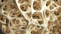
Osteoporosis is characterized by bone loss that exceeds bone building, resulting in bones that are porous, less dense, and prone to fracture. Unless diagnosed earlier, this condition is frequently confirmed only after a fracture or, sadly, after multiple fractures occur.2 Beyond its socioeconomic toll on the health-care system and families, osteoporosis drives severe clinical outcomes, including skeletal fragility, loss of autonomy, and heightened mortality following a major fracture.4
A silent killer
Osteoporosis, like hypertension, is often described as a “silent disease,” because it is typically asymptomatic until a fracture occurs or it is confirmed through bone screening.2,4 Like hypertension, it is also dubbed a silent killer because the mortality rate is high, especially in those with hip fractures. In the United States, approximately 300,000 patients will experience a hip fracture annually, and of these, a quarter will die in the following year, another quarter will step down from the hospital to skilled nursing and never return home, and the remaining half will never regain prior function or mobility.4
Peak bone mass
Osteoporosis is often considered a condition of aging; however, biological factors that influence its development begin much earlier in life. Leading experts and osteoporosis organizations note that adolescence is a pivotal period for bone development because a substantial proportion of peak bone mass (PBM) is accumulated during these years.5
According to Hereford et al.,5 PBM is the maximum amount of bone a person accumulates by the end of skeletal maturation, typically at around 30 years old. Bone mass increases rapidly throughout childhood and adolescence, with approximately 40%–60% of adult bone mass accrued during puberty. Individuals who achieve a higher PBM in youth can be better protected against osteoporosis and fractures later in life.
PBM results from the interaction of genetics, hormones, nutrition, physical activity, and overall health during growth and development. During adolescence, bone formation exceeds bone resorption, allowing substantial gains in bone mineral content and bone density. The pubertal growth spurt is especially important because bone mass accumulates rapidly during this period. After PBM is reached in early adulthood, bone mass ideally remains relatively stable until mid to late adulthood, when age-related bone loss begins.5 Maximizing bone accumulation during these years provides a larger “bone bank” that can help offset the bone loss many individuals encounter during their lifetime.6
PBM is shaped by both nonmodifiable factors (such as genetics, gender, race, ethnicity, and pubertal timing) and modifiable factors (including nutrition, physical activity, body weight, smoking, certain medications, and chronic health conditions). In males, higher body weight, greater muscle mass, and hormone levels, particularly testosterone, play a significant role in bone formation. Males can then achieve greater PBM than most females.5
Women and bone density
Bone homeostasis in women is orchestrated through the coordinated actions of sex hormones, including estrogen, progesterone, and testosterone, which regulate osteoblast and osteoclast activity to maintain skeletal growth, remodeling, and mineral balance. Estrogen preserves bone density by slowing its breakdown, testosterone contributes to bone formation and maintenance, and progesterone has been proposed to support osteoblastic bone formation.7 Subclinical bone loss—frequently classified as osteopenia—can begin decades before menopause during the premenopausal years.8 Driven by hormonal illustrations—most notably the decline in estradiol, the primary form of estrogen—bone density begins to decrease during perimenopause and continues to decline throughout menopause and beyond. Because this process occurs gradually and without noticeable symptoms, significant skeletal changes often go undetected until a more advanced stage is reached.9
Screening and diagnosis
Dual-energy x-ray absorptiometry (DXA or DEXA) is considered the gold standard for osteoporosis screening by measuring bone mineral density (BMD) and diagnosing osteoporosis. In the United States, many clinicians follow osteoporosis guidelines established by the U.S. Preventive Services Taskforce (USPSTF), which recommends a DEXA scan for women at age 65. If a woman has one or more risk factors for fracture—e.g., low body weight, smoking, parental hip fracture, or excess alcohol use—clinicians can prescribe the scan for those younger than age 65.10 Screening later in life often yields a diagnosis only after frailty has already taken root. Basic science strongly supports the notion that current screening guidelines must be revised and initiated at earlier ages for effective risk mitigation. In alignment with preventive medicine, the goal is to screen early, intercepting the disease before it manifests.
The most commonly evaluated skeletal sites during the DEXA scan are the lumbar spine (L1-L4), total hip, and femoral neck, because measurements obtained at these sites are used for osteoporosis diagnosis and fracture risk assessment. DEXA results are reported as a T-score, which compares an individual’s BMD with that of a healthy young adult reference population (20- to 29-year-olds) of the same sex and expresses the difference as standard deviations from the young-adult mean. The T-score expresses the number of standard deviations above or below this young-adult reference mean. A higher negative value indicates lower bone density and increased risk of fracture.11,12
Classification of bone mineral density according to T-score13:
- Normal: T-score -1.0 or higher
- Osteopenia (low bone mass): T-score between -1.0 and -2.5
- Osteoporosis: T-score of -2.5 or lower
- Severe or established osteoporosis: T-score -2.5 or lower in the presence of a fragility fracture
These classifications help clinicians assess fracture risk and guide preventive and therapeutic interventions.
Although BMD is an important determinant of bone strength, bone quality also contributes substantially to skeletal integrity. Think of bone quality as the collagen microarchitecture that serves as the scaffold for mineralization and the bone’s ability to absorb forces and resist fracture. Because DEXA measures BMD, it does not assess the aspects of bone quality. As a result, individuals with similar BMD measurements may have different fracture risks.14
Bone health and dentistry
Although osteoporosis is largely preventable, its asymptomatic nature, restrictive US screening guidelines, and lack of awareness leads to delayed diagnosis and intervention, resulting in significant clinical and economic burdens. Dental professionals are uniquely positioned to be part of the solution.15 This three-part educational series bridges that gap by moving dental clinicians into the forefront of early screening, education, and prevention, ultimately supporting their personal health and the health of their patients, peers, family, and friends.
Dental professionals are important members of the interprofessional health-care team. Through biannual dental hygiene visits and trusted relationships with patients, we are uniquely positioned to identify risk factors and contribute to improved health outcomes through the medical history review, oral examination, and dental radiographic assessments.16,17
A comprehensive medical history review can provide valuable insights into a patient's risk for bone conditions like osteoporosis. Important considerations can include things such as family history of osteoporosis or fracture, tobacco use, excessive alcohol consumption, perimenopause and menopause symptoms, and chronic corticosteroid use. Physical findings may also be suggestive of underlying skeletal concerns.
During the dental examination, clinicians may observe findings that have been associated with low bone mineral density, including increased alveolar bone loss, reduced trabecular bone density, and greater clinical attachment loss. These findings are not diagnostic of osteoporosis but may warrant consideration of the patient's overall osteoporosis risk and need for medical evaluation.18,19
The maxilla and mandible are large bones that continuously remodel throughout life. Like the hip and spine, these bones undergo ongoing bone turnover. Dental radiographs may reveal decreased mandibular cortical width, alterations in trabecular bone pattern, or increased alveolar bone loss, all of which have been investigated as potential indicators of low systemic BMD.18,19 While dental radiographs cannot diagnose osteoporosis, they may provide important clues that warrant further evaluation.
As dental providers, our care goes well beyond the oral cavity.17 By recognizing the early red flags during routine dental visits and conducting thorough medical history reviews, we can bridge the diagnostic divide for our patients. The oral cavity truly serves as a critical window to systemic health, empowering us to look beyond the teeth and gums. When clinical findings and systemic risk factors raise concern for low bone density, a referral for osteoporosis screening can narrow the care gap, turning early clinical awareness into proactive prevention and averting life-altering fractures.
True prevention, however, requires looking deeper into the foundational drivers of skeletal wellness. In part two of this series, we will explore comprehensive strategies for osteoporosis prevention in conjunction with optimizing women's health and longevity. See you next read!
Editor’s note: This article first appeared in Clinical Insights newsletter, a publication of the Endeavor Business Media Dental Group. Read more articles and subscribe.
References
- Salari N, Ghasemi H, Mohammadi L, et al. The global prevalence of osteoporosis in the world: a comprehensive systematic review and meta-analysis. J Orthop Surg Res. 2021;16(1):609. doi:10.1186/s13018-021-02772-0
- What is osteoporosis and what causes it. Bone Health & Osteoporosis Foundation. 2026. https://www.bonehealthandosteoporosis.org/patients/what-is-osteoporosis/
- Osteoporosis fast facts. Bone Health & Osteoporosis Foundation. 2026. https://www.bonehealthandosteoporosis.org/wp-content/uploads/Osteoporosis-Fast-Facts-2.pdf
- Facts about bone health. Bone Health & Osteoporosis Foundation. https://www.bonehealthpolicyinstitute.org/bone-facts
- Hereford T, Kellish A, Samora JB, Nichols LR. Understanding the importance of peak bone mass. J Pediatr Soc North Am. 2024;7:100031. doi:10.1016/j.jposna.2024.100031
- Bachrach LK. Acquisition of optimal bone mass in childhood and adolescence. Trends Endocrinol Metab. 2001;12(1):22-28. doi:10.1016/s1043-2760(00)00336-2
- Compston JE. Sex steroids and bone.Physiol Rev. 2001;81(1):419-447. doi:10.1152/physrev.2001.81.1.419
- Lo JC, Burnett-Bowie SAM, Finkelstein JS. Bone and the perimenopause. Obstet Gynecol Clin North Am. 2011;38(3):503-517. doi:10.1016/j.ogc.2011.07.001
- Streicher C, Heyny A, Andrukhova O, et al. Estrogen regulates bone turnover by targeting RANKL expression in bone lining cells. Sci Rep. 2017;7(1):6460. doi:10.1038/s41598-017-06614-0
- Kahwati LS, Kistler CE, Booth G, et al. Screening for osteoporosis to prevent fractures: a systematic evidence review for the US Preventive Services Task Force.JAMA. 2025;333(6):509-531. doi:10.1001/jama.2024.27154
- Punda M, Zhang-Yin J, Erba PA, et al. Dual-energy x-ray absorptiometry (DXA) in practice: a clinical centre survey endorsed by the European Association of Nuclear Medicine (EANM). Eur J Nucl Med Mol Imaging. 2025;52(4):1237-1241. doi:10.1007/s00259-024-07050-9
- Siris ES, Adler R, Bilezikian J, et al. The clinical diagnosis of osteoporosis: a position statement from the National Bone Health Alliance Working Group. Osteoporos Int. 2014;25(5):1439-1443. doi:10.1007/s00198-014-2655-z
- Faukner KG. The tale of the T-score: review and perspective. Osteoporos Int. 2005;16(4):347-352. doi:10.1007/s00198-004-1779-y
- Banks KP, Farrell MB, Gunther RS, McWhorter NE, Byerly DW, Peacock JG. Improving DXA quality by avoiding common technical and diagnostic pitfalls: part 1. J Nucl Med Technol. 2023;51(3):167-175. doi:10.2967/jnmt.122.264885
- Mills A, Berlin-Broner Y, Levin L. Improving patient well-being as a broader perspective in dentistry. Int Dent J.2023;73(6):785-792. doi:10.1016/j.identj.2023.05.005
- Witter D. Dentist visits predict wellbeing across income groups. Gallup. July 16, 2025. https://news.gallup.com/poll/692099/dentist-visits-predict-wellbeing-across-income-groups.aspx
- Gambhir RS. Primary care in dentistry – an untapped potential. J Family Med Prim Care. 2015;4(1):13-18. doi:10.4103/2249-4863.152239
- Zhu L, Zhou C, Chen S, et al. Osteoporosis and alveolar bone health in periodontitis niche: a predisposing factors-centered review. Cells. 2022;11(21):3380. doi:10.3390/cells11213380
- Alhumaidan AA, Elakel A. Systemic bone loss and periodontal disease: an updated review of a bidirectional association. Dent J. 2026;14(1):70. doi:10.3390/dj14010070
About the Author
 Kristin Evans, BS, RDH
Kristin Evans, BS, RDH
Kristin Evans, BS, RDH, is a nationally recognized dental educator with over 30 years of clinical experience. Known as “The pH RDH,” she specializes in pH balance, bone health, hormones, and the oral-systemic connection. Kristin delivers engaging, evidence-based education that bridges science and real-world application. Her dynamic style and deep knowledge empower dental teams to elevate outcomes, embrace prevention, and understand their vital role in improving whole-body health through personalized, integrative care.
Lori Stockert, DHSc, RDH, RDN
Lori Stockert, DHSc, RDH, RDN, is a licensed dental hygienist in Pennsylvania, New Jersey, and Australia, and a registered dietitian nutritionist in Pennsylvania. With more than 25 years of clinical dental hygiene experience, she has built a career spanning private practice, hospital-based care, and the pharmaceutical industry. Lori recently earned her Doctor of Health Sciences degree from Eastern Virginia Medical School. She is passionate about advancing preventive health care, promoting the connection between oral and overall health, and improving patient outcomes.