Squamous cell carcinoma: A pathology case report follow-up
Remember that case I posted about the pathology on the right posterior lateral border of the tongue that had some “meat” to it? The lesion was small, gave the patient zero discomfort, and would have gone on to be something much worse had we not caught it when we did. What was the definitive? Well, as suspected, it was cancer—squamous cell carcinoma (SCC) to be exact.
Refreshers regarding pathology are always good for us, so take a quick second and read up on this type of cancer that is more common than you think. Chances are, you’ve diagnosed this before; if you haven’t, keep looking because at some point in your career, you will.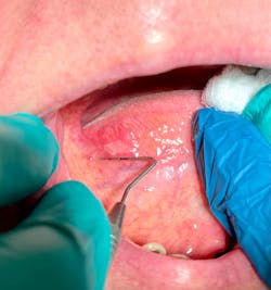
- What is SCC? It’s an end-stage alteration in stratified squamous epithelium, beginning as an epithelial dysplasia until the dysplastic epithelial cells breach the basement membrane and invade the connective tissue.
- Another common name is epidermoid carcinoma.
- SCC represents 3% of all cancer in males, 2% in females.
- The survival rate is 50%.
- SCC is the most common malignant neoplasm of the oral cavity, representing approximately 90% of all oral cancers.
- Etiologic factors: tobacco habits, alcohol consumption, viruses, actinic radiation, immunosuppression, nutritional deficiencies, preexisting diseases, and chronic irritation.
Related reading:
- Hoping my gut is wrong on this pathology case
- Would your progress notes hold up during a litigation case or state board hearing?
- Dental sealants: I love to hate ’em
- The extraoral and intraoral dental exam process for complete patient health
- Most common sites: lower lip (35%), lateral borders of the tongue (25%), floor of the mouth (20%), soft palate (15%), gingival/alveolar ridge (4%), and buccal mucosa (1%).
- Most common presentations are as leukoplakia and erythroplakia. The more advanced lesions may first appear as a painless ulcer, tumorous mass, or a verrucous (papillary growth).
- The lesions may have few surface changes but typically appear as a firm, indurated area with a deep infiltration into the connective tissue.
- Invasion of the bone (maxillary and mandibular) can result in loosening or loss of teeth; can also cause paresthesia with nerve involvement.
- Metastasizes by invading the lymphatic vessels, then progresses to the general lymph nodes where the lesions lodge and continue to proliferate. Enlarged, firm, and fixed lymph nodes are an ominous clinical sign.
- Clinical staging of the head and neck using the TNM system (T = primary tumor; N = regional lymph node; M = distant metastasis). There is a specific system for the oral cavity.
- Treatment is by surgical excision, radiation therapy, and if extensive enough, chemotherapy.
- Later diagnosis gives a poorer prognosis, especially for lesions on the lateral border of tongue or floor of mouth (as these lesions are more commonly advanced when they are finally diagnosed).
Report from the oral surgeon
The oral surgeon gave this report for my patient:
A biopsy of the ventrolateral tongue was performed on 11.9.21. The histopathologic report is consistent with squamous cell carcinoma with the lesion being completely excised. No further treatment is indicated [with me] at this time other than regular monitoring. I will be referring him to an oncologist for additional follow-up.
On my end, the patient is seen every three months for periodontal maintenance, and I will monitor the site for any residual concerns.
What’s significant about this outcome?
Am I glad this case went as it did? Definitely. The outcome for this patient could have been vastly different had the pathology not been caught early. It takes less than 45 seconds to do a complete head and neck exam, and once you come across something that is a little off-key, take a picture! There are absolutely NO reasons for you NOT to have cameras in your operatories. If you don’t have an intraoral camera, read this. And don’t forget to put a periodontal probe next to the lesion for size referencing. Finally, follow-up! So often these pathology referrals get lost and forgotten!
Real, raw, and unafraid dentistry is the name of the game.
More pathology cases
- Pathology case: An almost-vague radiodense lesion
- An extreme case of oral herpes
- Oral pathology case: Venous blue lake mole—what?
- Pathology with an iPhone. Yes, it's really that simple!
Editor’s note: This article first appeared in Through the Loupes newsletter, a publication of the Endeavor Business Media Dental Group. Read more articles and subscribe to Through the Loupes.
Reference
- Sapp, JP. Eversole, LR. Wysocki, GP. Contemporary Oral and Maxillofacial Pathology. Mosby.; 1997:174-182.
About the Author

Stacey L. Gividen, DDS
Stacey L. Gividen, DDS, a graduate of Marquette University School of Dentistry, is in private practice in Montana. She is a guest lecturer at the University of Montana in the Anatomy and Physiology Department. Dr. Gividen has contributed to DentistryIQ, Perio-Implant Advisory, and Dental Economics. You may contact her at [email protected].